 Translate
HCM All Hearts Diagnostic Tools
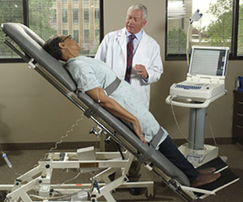
Echocardiogram

An echocardiogram is a test that uses sound waves to make images of your heart.
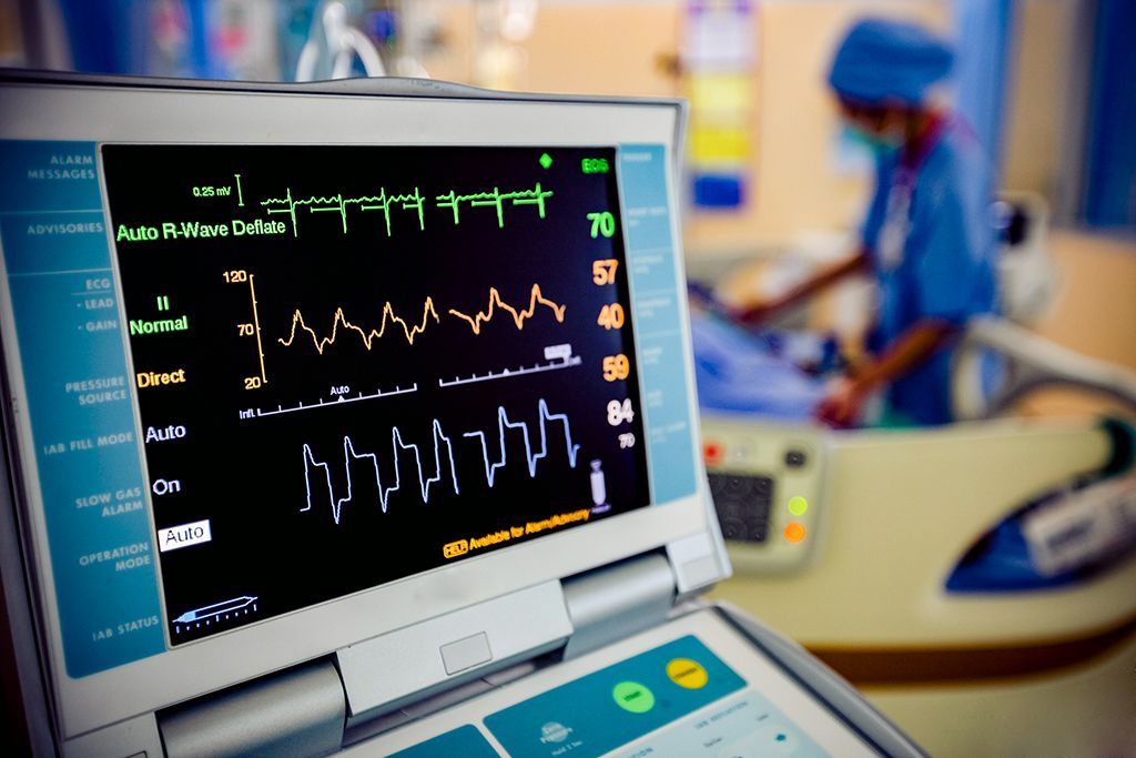
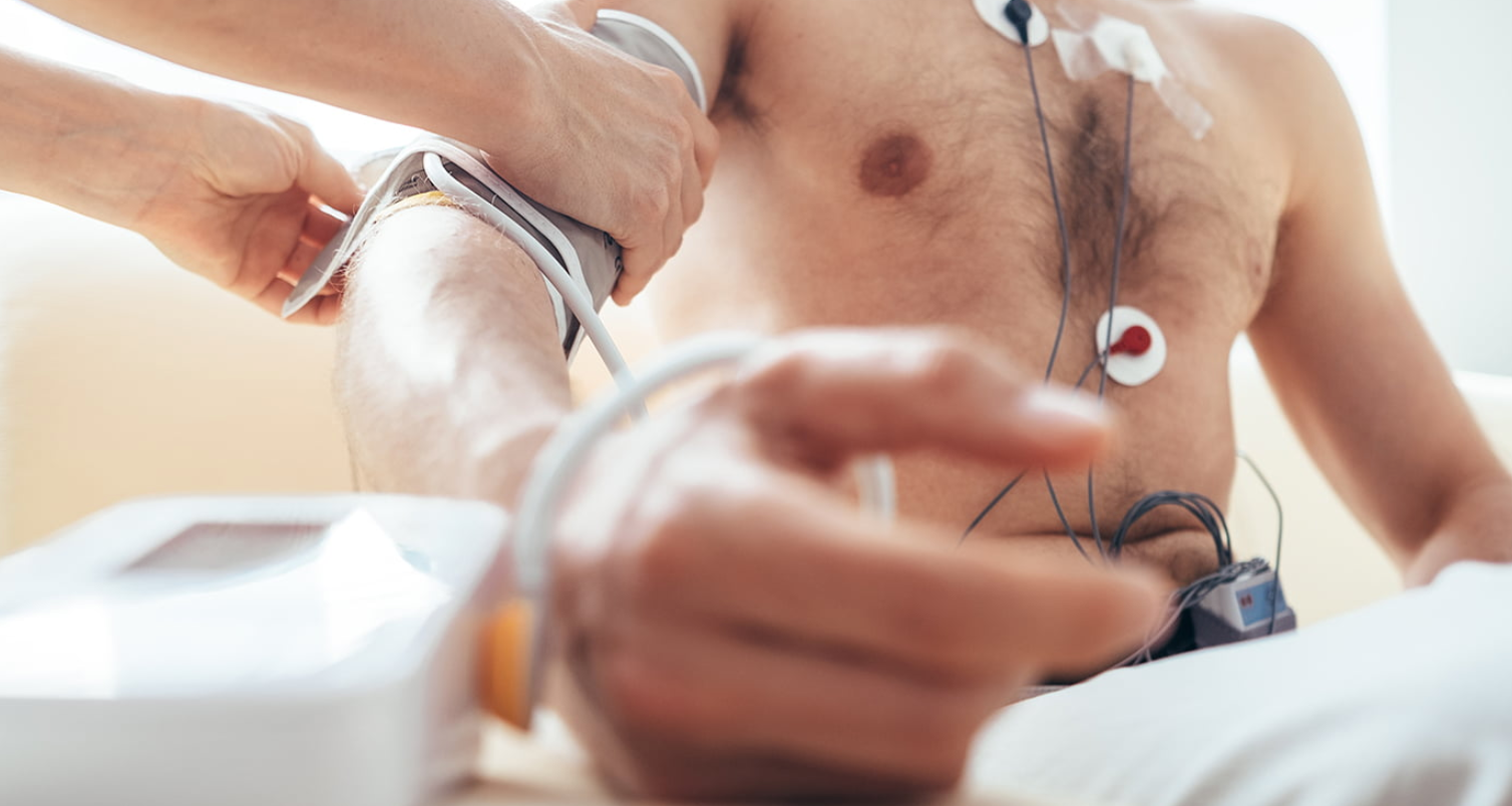
An echocardiogram (or echo) uses ultrasound to make images of your heart's chambers, valves, and walls. It can also image the major blood vessels attached to the heart. Some of these images are continuous, like movies. This means that an echo can be used to see things like how the valves move while the heart is pumping. Modern echocardiogram machines have the ability to use the Doppler effect. A familiar example of this effect is the difference in sound between a car moving toward you and one moving away. This allows your doctor to measure how blood moves in the different chambers and through the valves. Echos can be used to identify many heart disorders. The same basic technology is used in every other kind of medical ultrasound, including fetal ultrasounds.
Citations:
Mayo Clinic. (2018, October 04). Echocardiogram. Retrieved October 02, 2020, from https://www.mayoclinic.org/tests-procedures/echocardiogram/about/pac-20393856
American Heart Association. (2015, July 31). Echocardiogram (Echo). Retrieved October 02, 2020, from https://www.heart.org/en/health-topics/heart-attack/diagnosing-a-heart-attack/echocardiogram-echo
Cleveland Clinic. (2019, January 14). Echocardiogram. Retrieved October 02, 2020, from https://my.clevelandclinic.org/health/diagnostics/16947-echocardiogram
Related Diagnostic Tools