 Translate
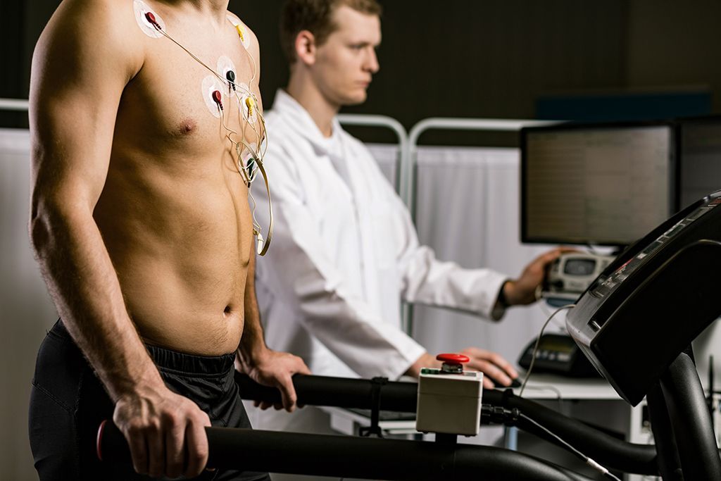
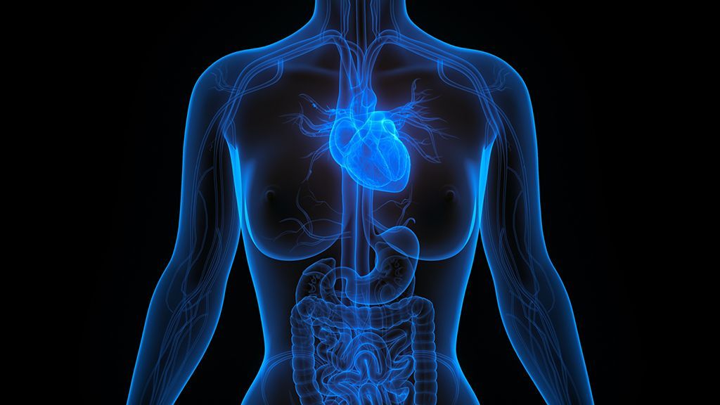
HCM All Hearts Diagnostic Tools
Transesophageal Echocardiogram (TEE)
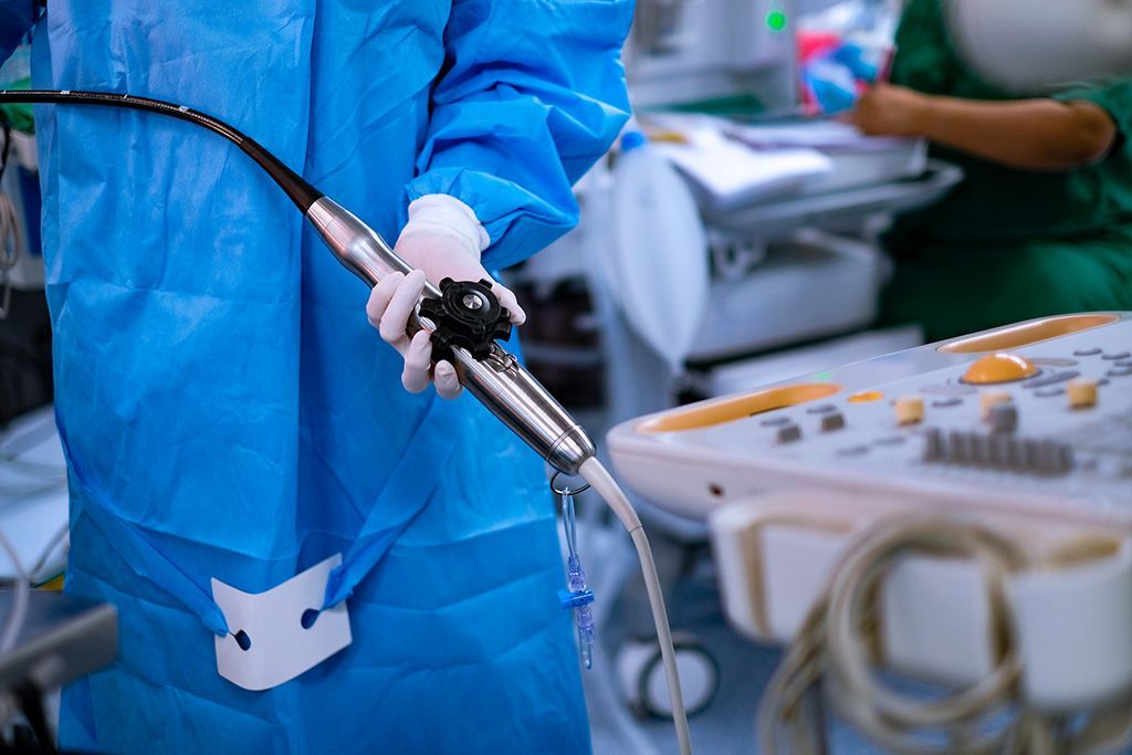
A transesophageal echocardiogram is a test that creates images of the heart. It is similar to a standard echocardiogram, but TEEs are done through the esophagus.
A transesophageal echocardiogram (TEE) provides real-time high-resolution images of the heart. It uses the same technology as a standard echocardiogram, but the measurements are taken by placing the transducer (wand) down the throat rather than across the chest and ribs.
If it is difficult to get a clear picture of your heart, or if your doctor wants a more in-depth image than what is produced by the standard transthoracic echocardiogram, a TEE can be used. A TEE may also be used if you have thick chest walls, are obese, are using a ventilator, or if there is an unremovable barrier between the transducer and your chest. TEEs are also used to see whether a clot (a stroke risk) is present in the atrium. In a TEE, the transducer is passed down your throat into your esophagus.
Your throat may be sore for a few hours after the TEE. Contact your doctor if this does not go away within a few days. You may also have trouble swallowing and have the urge to gag after the procedure.
Citations:
Mayo Clinic. (2018, October 04). Echocardiogram. Retrieved October 02, 2020, from https://www.mayoclinic.org/tests-procedures/echocardiogram/about/pac-20393856
American Heart Association. (2015, July 31). Transesophageal Echocardiography (TEE). Retrieved October 02, 2020, from https://www.heart.org/en/health-topics/heart-attack/diagnosing-a-heart-attack/transesophageal-echocardiography-tee
Johns Hopkins Medicine. (n.d.). Transesophageal Echocardiogram. Health. Retrieved October 02, 2020, from https://www.hopkinsmedicine.org/health/treatment-tests-and-therapies/transesophageal-echocardiogram
Related Diagnostic Tools